Small molecules self-organized in an orderly manner to enhance Raman signals
Raman spectroscopy is a powerful analytical technique that provides molecular fingerprints based on vibrational modes. However, its practical applications often face sensitivity limitations due to inherently weak Raman scattering signals. Traditionally, this challenge has been addressed using surface-enhanced Raman scattering (SERS) substrates, which rely on metallic nanostructures to amplify signals. Recently, a groundbreaking approach has emerged where small molecules self-organize into ordered stacks, enhancing Raman signals intrinsically without the need for external substrates. This phenomenon, known as stacking-induced intermolecular charge transfer-enhanced Raman scattering (SICTERS), opens new frontiers for non-invasive, high-resolution imaging and sensing. This article explores the mechanisms, materials, applications, and future prospects of this innovative self-assembly strategy to enhance Raman signals.
Fundamentals of Raman Spectroscopy and Signal Enhancement
Raman spectroscopy is based on the inelastic scattering of photons by molecular vibrations, providing detailed information about chemical structures. Despite its specificity, the Raman effect is intrinsically weak, making direct detection challenging, especially in dilute or complex biological samples.
To overcome this, signal enhancement techniques such as surface-enhanced Raman scattering (SERS) have been developed. SERS typically employs metallic nanostructures like gold or silver nanoparticles to amplify local electromagnetic fields, enhancing Raman signals by several orders of magnitude.
While SERS is effective, it often requires complex substrate preparation and can suffer from reproducibility and stability issues. Additionally, metallic substrates may interfere with biological environments, limiting in vivo applications. These challenges have motivated the exploration of alternative enhancement strategies that do not rely on external substrates.
The Concept of Small Molecule Self-Organization for Raman Enhancement
Self-organization is a process where molecules spontaneously arrange into ordered structures driven by non-covalent interactions such as π–π stacking, hydrogen bonding, and van der Waals forces. In the context of Raman enhancement, self-assembly of small molecules into ordered stacks can facilitate intermolecular charge transfer, leading to amplified Raman signals.
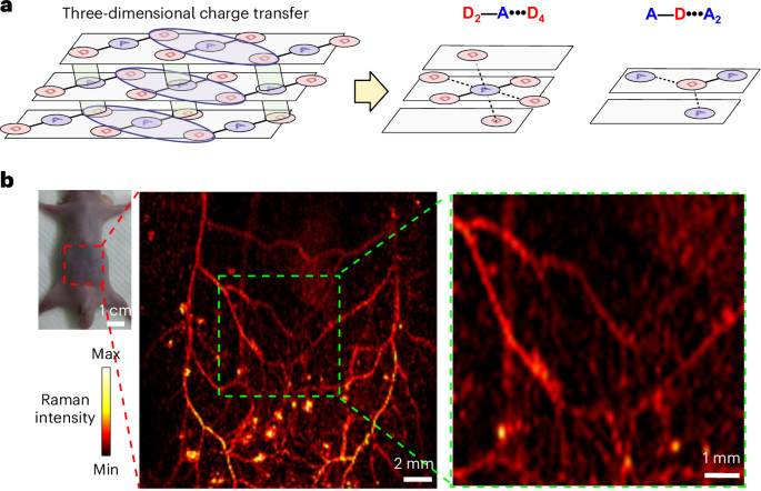
This stacking-induced intermolecular charge transfer-enhanced Raman scattering (SICTERS) effect leverages the intrinsic electronic interactions between adjacent molecules in the stack, creating new pathways for enhanced vibrational coupling and signal amplification.
Unlike traditional SERS, this approach eliminates the need for external metallic substrates, enabling substrate-free Raman probes. This intrinsic enhancement mechanism offers improved biocompatibility, stability, and reproducibility, making it highly attractive for biomedical imaging and sensing.
Mechanisms Behind Stacking-Induced Raman Signal Enhancement
At the molecular level, the ordered stacking of small molecules facilitates efficient intermolecular charge transfer. This charge transfer modifies the electronic environment of the molecules, increasing the polarizability and Raman scattering cross-section.
The π–π stacking interactions align the molecular orbitals, enabling delocalization of electrons across the stack. This delocalization enhances the coupling between electronic and vibrational states, amplifying Raman signals beyond what isolated molecules can achieve.
Furthermore, the self-stacking creates a nanoenvironment that stabilizes excited states and reduces non-radiative losses, contributing to stronger and more stable Raman signals. These combined effects underpin the SICTERS phenomenon, providing a robust platform for ultrasensitive Raman detection.
Materials and Molecular Design for Effective Self-Assembly
The choice of small molecules is critical for achieving effective self-assembly and Raman enhancement. Aromatic compounds with planar structures and strong π-conjugation, such as derivatives of pyrene, perylene, or porphyrins, are ideal candidates due to their propensity for π–π stacking.
Molecular modifications, including functional group substitution, can tune the stacking interactions and charge transfer efficiency. For example, introducing electron-donating or withdrawing groups can influence the electronic properties and optimize intermolecular interactions.
Additionally, designing molecules with amphiphilic characteristics can promote self-organization into well-defined nanostructures in aqueous environments, essential for biomedical applications. The rational molecular design thus enables control over assembly morphology and Raman enhancement performance.
Applications in Biomedical Imaging and Sensing
The substrate-free nature of self-organized small molecule Raman probes is particularly advantageous for in vivo biomedical imaging. These probes can penetrate biological tissues without introducing foreign metallic nanoparticles, reducing toxicity concerns.
High-resolution Raman imaging of lymphatic drainage and microvasculature has been demonstrated using SICTERS-active molecules, enabling non-invasive monitoring of physiological processes with exceptional spatial detail.
Furthermore, these probes facilitate real-time, label-free detection of biomolecules and pathological changes, offering promising diagnostic potential in cancer, inflammation, and metabolic disorders. Their stability and biocompatibility also support longitudinal studies in living organisms.
Advantages Over Conventional Raman Enhancement Techniques
Self-organized small molecule Raman probes circumvent many limitations of traditional SERS substrates, including batch-to-batch variability, substrate degradation, and complex fabrication processes.
The intrinsic enhancement mechanism promotes uniform and reproducible Raman signals, improving quantitative analysis reliability. Additionally, the absence of metal nanoparticles reduces background interference and potential cytotoxicity.
This approach also offers enhanced flexibility in probe design, allowing customization for specific molecular targets or imaging modalities. The substrate-free system simplifies experimental setups and broadens the scope of Raman spectroscopy applications.
Challenges and Future Perspectives in Small Molecule Self-Stacking
Despite its promise, the self-assembly approach faces challenges including controlling the uniformity and stability of molecular stacks in complex biological environments. Environmental factors such as pH, ionic strength, and competing biomolecules can influence assembly behavior.
Further research is needed to elucidate the detailed charge transfer dynamics within stacks and optimize molecular designs for maximal Raman enhancement. Advanced computational modeling combined with experimental spectroscopy can accelerate this understanding.
Looking forward, integrating self-assembled Raman probes with multimodal imaging platforms and developing stimuli-responsive assemblies could unlock new diagnostic and therapeutic applications. Collaborative efforts between chemists, physicists, and biologists will be vital to fully exploit this innovative technology.
Emerging Trends and Innovations in Raman Signal Enhancement
Recent innovations include hybrid systems combining self-assembled small molecules with nanostructured materials to synergistically enhance Raman signals while maintaining biocompatibility.
The development of dynamic self-assembly systems responsive to biological stimuli, such as enzymes or pH changes, is enabling targeted and controlled Raman imaging with high specificity.
Machine learning approaches are being employed to analyze complex Raman spectra obtained from self-assembled probes, enhancing signal interpretation and enabling rapid diagnostics. These trends highlight the evolving landscape of Raman spectroscopy powered by molecular self-organization.
Conclusion
The advent of small molecules self-organizing into orderly stacks to enhance Raman signals represents a paradigm shift in Raman spectroscopy. By harnessing intrinsic intermolecular charge transfer mechanisms, this substrate-free approach overcomes longstanding limitations of traditional enhancement techniques. The resulting ultrasensitive, biocompatible Raman probes open exciting avenues for non-invasive biomedical imaging and precise molecular sensing. While challenges in assembly control and mechanistic understanding persist, ongoing research promises to refine this technology further. Ultimately, self-assembled small molecule Raman probes are poised to become indispensable tools in chemical analysis, diagnostics, and beyond.
Originally reported by nature.com. Adapted for our readers.
Recent advances in machine perfusion technology are not only preserving donated livers but also making them biologically younger at a molecular level. This breakthrough…
What happens when human imagination, artificial intelligence, art and technology come together? From September 1 to 6, Samsung Electronics presented a special exhibition themed…